Persistent Right Aortic Arch (PRAA) in Cats

Learn about PRAA in cats, from early symptoms like regurgitation to advanced surgical treatments and recovery, backed by real clinical case studies and expert insights.
What you will read in this article
Introduction: Why Is Your Kitten Regurgitating After Solid Food?
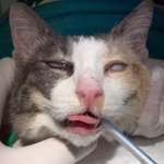
The joy of bringing home a new kitten can quickly turn into concern when you notice they are struggling to keep food down. Many owners observe that their kitten is energetic and has a voracious appetite, yet they remain smaller and thinner than their littermates. This classic scenario often points to a Vascular Ring Anomaly, specifically Persistent Right Aortic Arch (PRAA). While kittens do well on milk, the transition to solid food reveals a mechanical obstruction that prevents nourishment from reaching the stomach.
Understanding this condition is critical because it is not a simple digestive upset; it is an anatomical entrapment of the esophagus. Early intervention is the key to preventing permanent damage and ensuring a normal life for these feline patients. In this guide, we combine the latest veterinary research with real-world surgical insights to explain how we diagnose and cure this challenging condition.
What is Persistent Right Aortic Arch (PRAA) in Cats? (The Pathophysiology)
To understand Persistent Right Aortic Arch (PRAA), we must look at embryological development. During fetal growth, several pairs of aortic arches exist. Normally, the left fourth aortic arch develops into the permanent adult aorta, while the right one regresses. In cats with PRAA, this process is reversed: the right fourth aortic arch persists as the functional aorta. This anatomical “mistake” creates a trap for the esophagus at the level of the heart base.
The “ring” that constricts the esophagus is formed by three main structures: the persistent right aorta on the right, the pulmonary artery on the left and ventral side, and most importantly, the ligamentum arteriosum (the remnant of the ductus arteriosus) which crosses over the dorsal surface of the esophagus to connect the two. This tight band acts like a noose, preventing the esophagus from expanding when food passes through. As a result, the portion of the esophagus in front of the heart becomes severely dilated, a condition known as Megaesophagus.
The Embryological Error: How the Vascular Ring Forms
In the early stages of fetal development, the arterial system begins as a series of six pairs of aortic arches. Under normal conditions, the left fourth aortic arch persists to become the definitive adult aorta, while the right one regresses. However, in PRAA cases, a critical error occurs: the right fourth arch remains functional and the left one disappears.
This reversal would not be a problem on its own, but the Ductus Arteriosus (which becomes the Ligamentum Arteriosum after birth) still develops from the left sixth arch. This creates a lethal intersection: the aorta is now on the right of the esophagus, and the ligamentum travels from the left pulmonary artery to the right-sided aorta, passing directly over the dorsal aspect of the esophagus. This “vascular ring” effectively clamps the esophagus against the base of the heart, leading to the clinical signs we observe.
Common Symptoms and Clinical Signs: What Owners and Vets Should Watch For
The clinical presentation of PRAA is remarkably consistent. Symptoms typically emerge during weaning, as the kitten transitions from a liquid diet (milk) to semi-solid or solid kibble. While liquids can easily slide through the narrowed esophagus at the heart base, solid boluses of food get stuck. This leads to the most common sign: postprandial regurgitation. Owners often report that the kitten is always hungry and eats quickly, only to have the food come back up almost immediately. Over time, this lack of nutrient absorption leads to stunting and muscle wasting, making the affected kitten look much smaller than its healthy siblings.
Regurgitation vs. Vomiting: Understanding the Difference
It is vital to distinguish between regurgitation and vomiting. Vomiting is an active process involving abdominal contractions, retching, and often bile. In contrast, regurgitation in PRAA cats is passive and effortless. The food is expelled from the esophagus before it ever reaches the stomach, so it typically lacks the acidic smell of gastric juice and often maintains a tubular shape. Recognizing this distinction is the first step toward suspecting a vascular ring anomaly rather than a primary gastrointestinal disease.
The Risk of Aspiration Pneumonia: A Silent Threat
The most dangerous complication of PRAA is Aspiration Pneumonia. Because the esophagus is dilated and filled with undigested food (Megaesophagus), material can easily be inhaled into the lungs, especially during sleep or after an episode of regurgitation. Clinicians should look for signs like coughing, nasal discharge, fever, and lethargy. In many cases, it is the respiratory distress, rather than the digestive issue, that first brings the kitten to the veterinary clinic. According to the reference by Fetto, managing this pneumonia is often necessary before the patient is stable enough for anesthesia and surgery.
Diagnostic Imaging: How We Confirm PRAA
Confirming a diagnosis of PRAA requires imaging that goes beyond a standard physical exam. While the clinical history of a kitten regurgitating solid food is highly suggestive, we need to visualize the obstruction and its effects on the esophagus. Diagnostic imaging not only confirms the presence of a Vascular Ring Anomaly but also helps the surgical team rule out other causes of megaesophagus or esophageal foreign bodies.
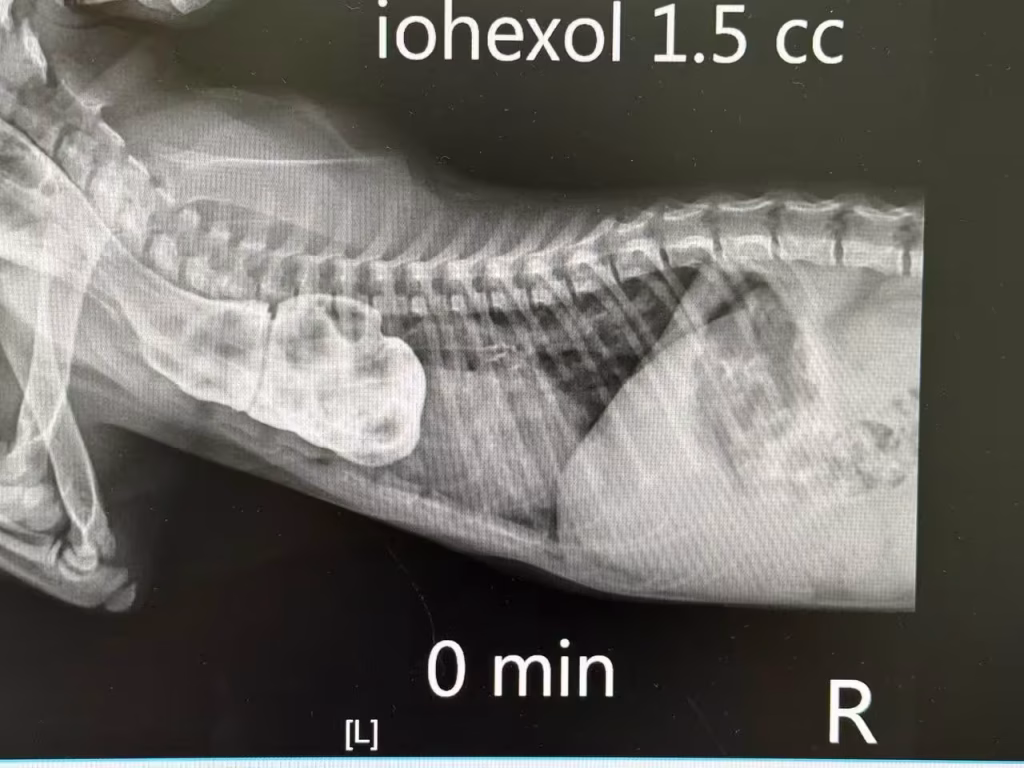
Barium Contrast Radiography: The Classic Megaesophagus View
The gold standard for initial diagnosis is Barium Contrast Radiography (an esophagram). By administering a liquid contrast agent, we can clearly see the esophagus dilated cranial (in front) to the heart base, followed by a sudden narrowing at the site of the vascular ring. According to Merck Veterinary Manual, a highly specific sign of PRAA is the rightward deviation of the trachea on a dorsoventral (DV) view, caused by the abnormal position of the aortic arch.
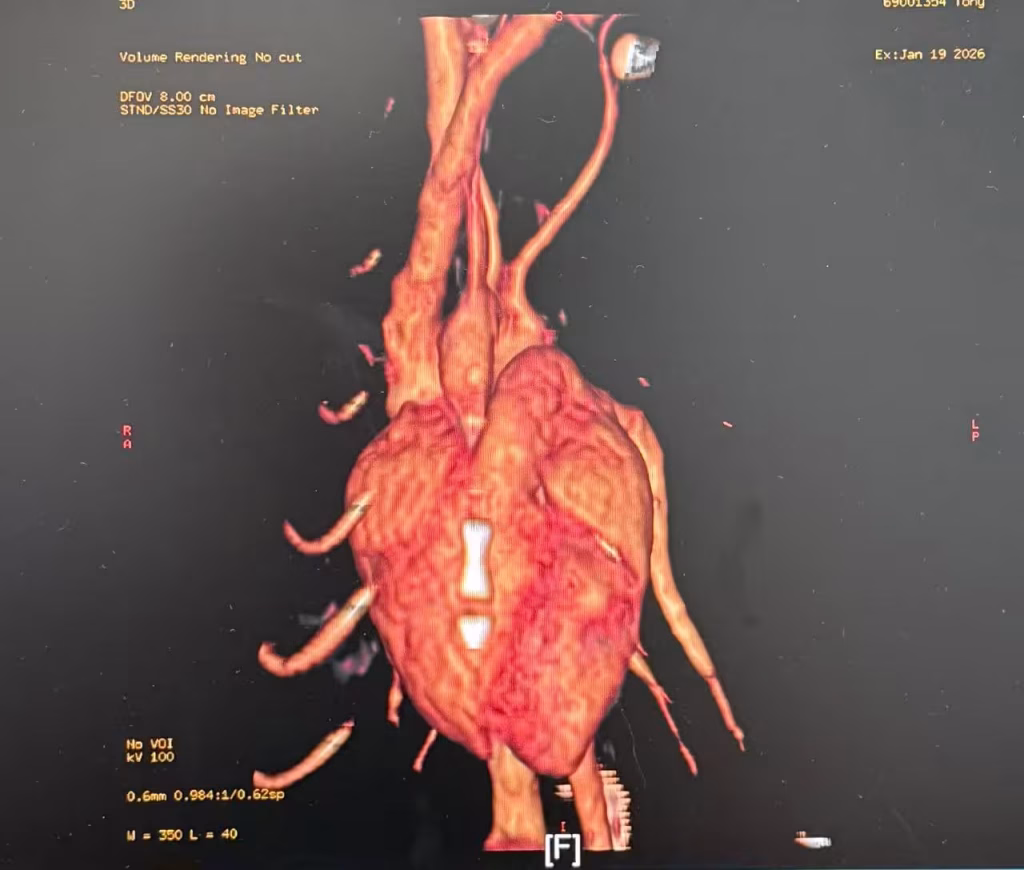
The Role of CT Angiography in Surgical Planning
While radiography confirms the obstruction, CT Angiography provides a 3D roadmap for the surgeon. In cats, vascular anomalies can be complex, sometimes involving an aberrant subclavian artery. CT allows us to identify the exact vessels forming the ring and their relationship to the esophagus and trachea. This precision, as highlighted in the Bascuñán study, is vital for choosing the correct surgical approach and avoiding life-threatening intraoperative hemorrhages.
Surgical Management: Life-Saving Procedures (Case Study Analysis)
Surgery is the only definitive treatment for PRAA. The primary goal is to break the vascular ring and release the constricted esophagus. Without surgical intervention, the prognosis is poor due to progressive starvation and recurrent pneumonia. Today’s case study provides a perfect example of how precision and anatomical knowledge come together in the operating theater to save a kitten’s life.
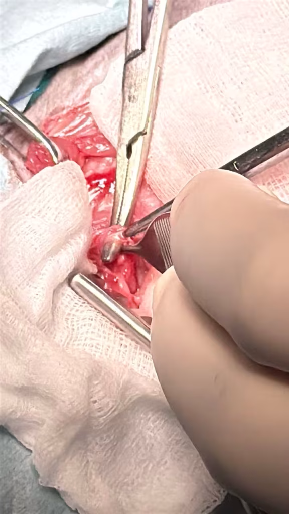
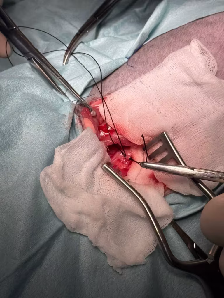
Intercostal Thoracotomy: The Gold Standard Approach
The most common approach, as seen in our current case, is a left-sided fourth intercostal thoracotomy. This provides direct access to the ligamentum arteriosum. Once the thoracic cavity is opened, the surgeon must carefully retract the lungs to visualize the mediastinum. In the provided surgical images, the clear exposure of the heart base and the overlying vessels demonstrates the effectiveness of this classic approach for ensuring a safe ligation.
Thoracoscopic Correction (VATS): The Future of Feline Surgery
While open surgery is the standard, Video-Assisted Thoracoscopic Surgery (VATS) is gaining popularity in feline medicine. As discussed in the Radlinsky reference, VATS offers superior magnification and lighting, which is crucial given the small size of a cat’s thorax. This minimally invasive technique leads to significantly less post-operative pain and faster recovery times, although it requires specialized equipment and advanced technical skills.
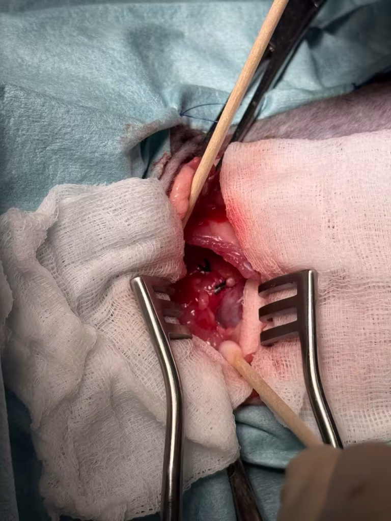
Intraoperative Challenges: Freeing the Esophagus
Cutting the ligamentum is only half the battle. The real challenge, visible in the surgical photos, is the meticulous dissection of periesophageal fibrous bands. These bands often persist even after the main ring is broken, continuing to constrict the esophagus. Surgeons often use a Foley catheter or an orogastric tube during the procedure to identify the narrowing and ensure the esophagus is fully mobile. Care must be taken to avoid damaging the Vagus and Phrenic nerves, which run in close proximity to the surgical site.
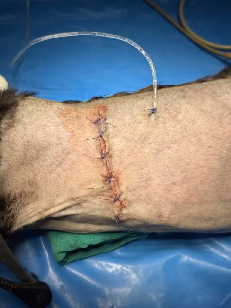
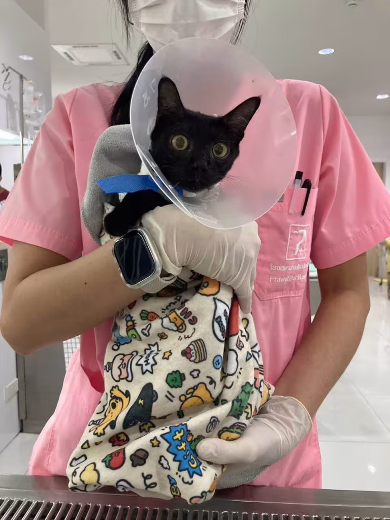
Post-Operative Care and Long-Term Prognosis
The success of a PRAA surgery is not solely determined in the operating room. Post-operative management is a marathon, not a sprint. While the mechanical obstruction is removed, the esophagus often remains dilated and has poor motility. Immediate post-op care focuses on pain management, monitoring for chest tube complications (if used), and preventing further episodes of aspiration. The long-term goal is to transition the kitten to a lifestyle that minimizes the risk of food stagnation in the esophagus.
Feeding Strategies: Living with a Recovering Esophagus
Feeding after PRAA surgery requires a “gravity-assisted” approach. Kittens should be fed in an upright position (using a Bailey chair or holding them vertically) to allow gravity to push food into the stomach. The consistency of the food is also crucial; some cats do better with a liquid slurry, while others handle small, “meatball” shaped soft food better. Owners must continue this elevated feeding for weeks, or sometimes indefinitely, depending on the degree of esophageal recovery.
Will the Megaesophagus Ever Resolve? (What Science Says)
One of the most common questions owners ask is whether the esophagus will return to normal size. Science offers a cautious answer. According to the study by Muldoon et al., while clinical signs like regurgitation improve significantly in over 90% of cases, radiographic megaesophagus often persists to some degree. The esophagus may never regain full motor function, but most cats can lead a happy, healthy life with proper dietary management. Early surgical intervention (before the kitten reaches 4 months of age) is the most significant factor in achieving better esophageal remodeling.
Frequently Asked Questions (FAQ) About PRAA in Cats
1. Is PRAA a hereditary condition in cats?
Yes, it is widely believed to be a congenital abnormality with a complex hereditary basis. According to the Fetto reference, it involves recessive genes. Therefore, it is strongly recommended that affected cats and their littermates should not be used for breeding.
2. Can PRAA be managed without surgery?
While small meals and upright feeding can temporarily help, surgery is the only definitive cure. Without it, the cat will suffer from chronic malnutrition and is at high risk of fatal Aspiration Pneumonia. Medical management alone is generally not recommended for long-term survival.
3. At what age should the surgery be performed?
Ideally, as soon as the condition is diagnosed, typically between 2 to 6 months of age. Early intervention (ideally before 4 months) provides the best chance for the esophagus to regain some of its function, as highlighted in the Muldoon study.
4. What is the success rate of the surgery?
The short-term survival rate is excellent, often exceeding 90%. However, long-term success depends on the severity of the megaesophagus before surgery. Most cats will show significant clinical improvement, though some dietary modifications may be needed permanently.
Conclusion: The Take-Home Message
Persistent Right Aortic Arch is a challenging but treatable condition. The journey from a struggling kitten to a healthy cat depends on early diagnosis and precise surgical intervention. As demonstrated in today’s surgical case, the liberation of the esophagus from its vascular entrapment offers these patients a second chance at life. While post-operative dietary management is often required, the majority of feline patients go on to live a normal, high-quality life.
Scientific References
- Bascuñán, A., et al. Vascular ring anomalies in cats: 20 cases (2000–2018)
- Radlinsky, M. Thoracoscopy in the cat: An up-and-coming diagnostic and therapeutic procedure
- Muldoon, M. M., et al. Long-term results of surgical correction of persistent right aortic arch in dogs: 25 cases (1980–1995)
- Fetto, J. Understanding Vascular Ring Anomaly
- Merck Veterinary Manual – Vascular Malformations in Animals (Circulatory System)
Clinical Image Credit:
We would like to express our sincere gratitude to Dr. Wijit Sutthiprapa (DVM, MS, Dip. TBVS) for providing the clinical images used in this article. His contribution as a specialist in the field enhances the educational value of this guide for pet owners and veterinary students alike.