Canine Hepatoid Gland Tumors: A Complete Guide to Diagnosis and Treatment

Discovering a mass around a dog’s anus can be deeply concerning for any pet owner, especially when your pet is in its senior years. Perianal gland tumors, also known as Hepatoid Gland Tumors, are among the most common neoplasms in older male dogs. Early diagnosis through cytology and prompt surgical intervention can significantly improve an animal’s quality of life. In this article, we meticulously analyze this condition based on the authoritative Merck Veterinary Manual and provide a detailed analysis of a real-life surgical case involving an invasive mass in a 15-year-old dog to answer all your questions.
What you will read in this article
What are Hepatoid or Perianal Gland Tumors? (Origin and Nature)
Hepatoid glands are modified sebaceous glands found primarily in the skin surrounding the anus of dogs. The term “Hepatoid” is derived from their microscopic resemblance to liver cells (Hepatocytes).
According to the Merck Veterinary Manual, these tumors are part of the perianal gland tumor family and manifest in three primary forms:
- Adenoma: The most common form, which is benign and typically slow-growing.
- Epithelioma: Tumors with more immature cells that possess the potential for invasive growth into surrounding tissues.
- Adenocarcinoma: The malignant or cancerous form, which is 10 times more common in male dogs than females and can metastasize to regional lymph nodes.
A crucial point for every dog owner is that the growth of these glands is heavily influenced by sex hormones, particularly testosterone. Consequently, intact senior male dogs are at the highest risk. Even crossbreeds (like our current case) may face this significant challenge in their later years if not castrated.
Comparative Table of Hepatoid Gland Tumors
| Feature | Adenoma (Benign) | Epithelioma (Intermediate) | Adenocarcinoma (Malignant) |
| Growth Rate | Slow | Moderate | Fast and Invasive |
| Clinical Appearance | Small, solitary nodules | Firm, infiltrative masses | Large, ulcerated, infected masses |
| Response to Castration | Excellent (up to 95%) | Partial | Poor or None |
| Metastatic Potential | None | Very Rare | High (to lymph nodes) |
| Gender Prevalence | Intact males | Males | Both (More dangerous in females) |
Case Report: Surgical Challenge of an Invasive Mass in a 15-Year-Old Dog
This section explores a clinical case of a 15-year-old male crossbreed dog presented with multiple firm masses in the perianal region. Managing such a case in a geriatric patient requires careful pre-anesthetic evaluation. The dog, identified with DEA1- blood type, exhibited multifocal growths around the anal sphincter. This case is a classic example of how hepatoid gland tumors progress in intact senior dogs, demanding immediate and precise surgical intervention.
Demographic information and clinical status of the case: patient profile and physical examination
| Pet Name | Lucky |
| Age | 15 Years and 19 Days |
| Gender | Male (Intact) |
| Breed | Crossbreed |
| Blood Type | DEA1 Negative |
| Mass Margin | Invasive |
| Content | Seropurulent |
| Consistency | Firm |
| Distribution | Multifocal |
Pre-operative Analysis: Multifocal and Invasive Masses
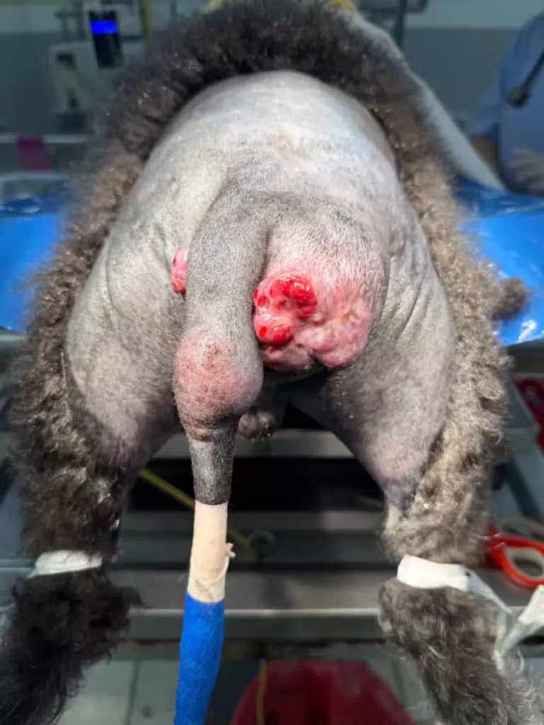
Pre-operative images of this case reveal masses with invasive margins that significantly distorted the perineal and perianal anatomy. Clinical examination confirmed a very firm consistency, which is often an indicator of potential malignancy. Additionally, the presence of seropurulent discharge pointed toward severe inflammation and secondary infection at the tumor site. As shown in the images, the tumor was multifocal, involving not only the skin surface but also infiltrating deeper tissues. The extent of this involvement necessitated a wide surgical excision with a clear safety margin.
Clinical Signs: When to Worry About Anal Masses in Dogs?
Many dog owners initially mistake hepatoid gland tumors for more common issues like anal sac inflammation or even constipation. According to the Merck Manual, these tumors typically appear as firm, round, single or multiple nodules under the skin around the anus.
Symptoms you should take seriously include:
- Visible Prominent Masses: Nodules that can range from a few millimeters to several centimeters in diameter.
- Ulceration and Bleeding: Larger tumors (like the one seen in our case) often become ulcerated and may show infected or bloody discharge.
- Changes in Defecation: Straining during bowel movements (Tenesmus) due to the pressure the mass exerts on the anal canal.
- Excessive Licking: The dog may constantly lick the area under the tail and around the anus due to discomfort, pain, or irritation.
- Swelling and Redness: In invasive cases, the entire perineal region may appear inflamed and painful.
Diagnostic Tools: Cytology (FNA) vs. Tissue Biopsy
Accurate identification of a perianal mass is the first step toward successful treatment. In our clinical case, Cytology or Fine Needle Aspiration (FNA) was utilized. The lab report for this 15-year-old dog confirmed the presence of epithelial cells with invasive characteristics, suggesting a spectrum from hepatoid epithelioma to adenocarcinoma.
Details of cytology findings: microscopic analysis of FNA sampling
| Cytological Parameter | Findings Extracted from Lab Report |
| Cellular Arrangement | Cohesive clusters of large polygonal cells |
| Nuclear Status | Round to oval nuclei with hyperchromatic chromatin |
| Malignancy Indices | Altered N:C ratio and presence of Nuclear molding |
| Size Variation | Mild to moderate anisokaryosis and anisocytosis |
| Presumptive Diagnosis | Hepatoid gland epithelioma to adenocarcinoma |
| Final Recommendation | Histopathological confirmation via tissue biopsy |
According to the Merck Veterinary Manual, differential diagnosis in this region is critical:
- Cytology: A quick, non-invasive method to confirm the hepatoid origin of the mass. However, distinguishing between a benign adenoma and a low-grade adenocarcinoma can be challenging through cytology alone.
- Tissue Biopsy (Histopathology): Merck emphasizes that definitive diagnosis of malignancy requires evaluating the tissue architecture through histopathology.
- Differential Diagnosis: It is essential to differentiate hepatoid gland tumors from anal sac adenocarcinomas, as the latter are far more aggressive and often associated with hypercalcemia.
Given the patient’s age and the invasive findings on cytology, the surgeon proceeded directly with wide surgical excision.
Differential Diagnosis: Perianal vs. Adenocarcinoma
It is common for pet owners and veterinary students to use Perianal Gland Tumors and Hepatoid Gland Adenocarcinoma interchangeably, but they represent different clinical levels:
- Perianal/Circumanal Gland Tumor: This is a general term describing any neoplasm arising from the glands surrounding the anus. It encompasses both benign (Adenoma) and malignant (Adenocarcinoma) forms.
- Hepatoid Gland Adenocarcinoma: This is a specific pathological diagnosis for the malignant form. It is characterized by invasive growth into underlying tissues and is often independent of testosterone levels, meaning castration alone will not resolve the mass.
In our 15-year-old case, based on the cytological report and the observed invasive behavior, the diagnosis shifted from a simple perianal tumor to a Hepatoid Adenocarcinoma, necessitating an aggressive surgical approach.
Comparison Table: General Terms vs. Specific Diagnosis
| Feature | Perianal / Circumanal Gland Tumor | Hepatoid Gland Adenocarcinoma |
| Scientific Definition | General term for any mass in this region | Specific diagnosis for a malignant tumor |
| Inclusions | Both Adenoma (benign) and Carcinoma | Only Adenocarcinoma (malignant) |
| Hormone Dependency | Usually testosterone-dependent | Often independent of sex hormones |
| Growth Rate | Can be slow and well-circumscribed | Rapid, invasive, and infiltrative |
| Surgical Goal | Mass removal and castration | Aggressive Wide Marginal Excision |
Why are Male Dogs More Prone to Hepatoid Gland Cancer?
One of the most striking characteristics of hepatoid gland tumors is their strong dependence on sex hormones. According to the Merck Veterinary Manual, these tumors are 10 times more common in intact male dogs than in females.
The reasons for this prevalence include:
- The Role of Testosterone: Hepatoid glands possess hormonal receptors. Testosterone, produced in the testes, directly stimulates the growth and proliferation of these gland cells. In senior dogs exposed to this hormone for years, the risk of cellular mutation and tumor formation increases significantly.
- The Effect of Estrogen: Interestingly, estrogen has an inhibitory effect on these glands. This explains why the disease is so rare in female dogs.
- Adenocarcinoma in Females: Merck notes that if a female dog or a neutered male develops this tumor, the likelihood of it being malignant (adenocarcinoma) is much higher, as the mass grew without hormonal stimulation, indicating a more aggressive nature.
In our 15-year-old case, being intact was the primary driver for the multifocal tumor growth. This is why the surgeon recommends castration alongside tumor removal to eliminate the hormonal source and prevent recurrence.
Treatment Options (Merck Ref): Surgery and Castration
Treating hepatoid gland tumors requires a dual strategy: removing the current mass and preventing future hormonal stimulation. According to the Merck Veterinary Manual, the most effective treatments include:
- Wide Surgical Excision: For tumors that appear invasive or multifocal (as in our case), removing the mass with a clear safety margin is vital. In our 15-year-old patient, the surgeon carefully excised the affected tissues to prevent residual tumor cells.
- Castration: This is the cornerstone of treatment for male dogs. Merck states that up to 95% of hepatoid adenomas regress or stop growing following castration. Even in malignant cases, neutering is essential to control the hormonal environment.
- Cryosurgery: For very small masses (less than 1 cm), freezing the tissue can be effective, but it is not suitable for advanced cases like the one in this report.
- Radiation Therapy: If the tumor is an adenocarcinoma and complete surgical removal is not possible due to proximity to the anal sphincter, radiation is recommended as an adjunctive therapy.
In our case, given the suspicious findings on cytology, the surgeon opted for wide excision and aggressive regional management to ensure the best possible outcome.
Surgical Technique and Perineal Reconstruction (Post-operative Analysis)
Surgery in the perineal region is exceptionally challenging due to its proximity to the anal sphincter and vital nerves. In our 15-year-old patient, the surgeon employed a Wide Marginal Excision technique. Since the masses were multifocal, a significant portion of the perianal skin had to be removed.
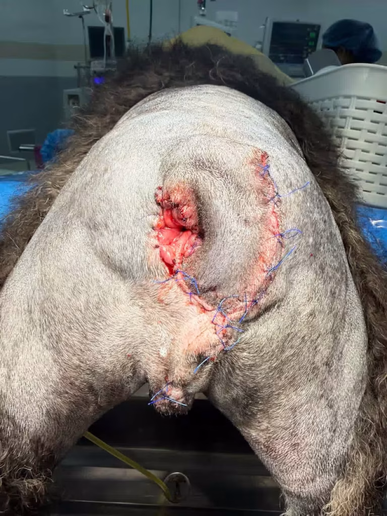
Key observations from the post-operative images:
- Preservation of Anal Sphincter Function: The surgeon’s top priority was protecting the external anal sphincter muscle to prevent fecal incontinence.
- Dead Space Management: After removing large tumors, a void is created under the skin. The surgeon utilized precise subcutaneous sutures to close this space and prevent fluid accumulation (seroma).
- Tissue Reconstruction: Due to the extensive tissue removal, advanced suturing techniques were used to appose the skin edges without excessive tension. High tension in this area can lead to suture line failure (dehiscence).
- Final Appearance: The immediate post-operative images show a clean, symmetrical suture line, demonstrating the surgeon’s skill in restoring the region’s natural anatomy.
Case Management Timeline
| Management Stage | Time / Status | Clinical Significance |
| Patient Admission | 09:28 AM | Initial stabilization and geriatric assessment |
| FNA Sampling | Jan 28, 2026 | Rapid cytological diagnosis before major surgery |
| Blood Compatibility | DEA1 Negative | Preparing for potential transfusion in senior patients |
| Surgical Procedure | Same Day | Wide marginal excision of multifocal masses |
| Post-Op Discharge | 16:44 PM | Stable vital signs and successful early recovery |
Post-operative Care and Disease Prognosis
Post-operative care for perianal tumors is just as critical as the surgery itself. In our 15-year-old patient, given the sensitive location and the dog’s age, careful recovery management was the key to success.
Key Post-operative Care Points:
- Elizabethan Collar (E-Collar): Consistent use of an E-collar is mandatory to prevent the dog from licking or chewing the sutures.
- Stool Management: Using stool softeners or specific diets prescribed by the vet is crucial to avoid constipation and reduce tension on the suture line during defecation.
- Hygiene: Keeping the surgical site clean and free of fecal contamination is essential to prevent secondary infections.
Prognosis According to Merck:
The prognosis depends largely on the histopathological type of the tumor:
- Adenoma (Benign): With successful surgery and castration, the prognosis is excellent, and the recurrence rate is very low.
- Adenocarcinoma (Malignant): According to the Merck Manual, the prognosis for malignant cases is “guarded.” If the tumor has metastasized to regional lymph nodes, management becomes more complex. However, in the case we analyzed, the aggressive wide excision aimed to provide the highest quality of life for the dog’s remaining years.
Pharmacological Management and Chemotherapy
According to the Merck Manual text in the provided records, malignant hepatoid gland adenocarcinomas are generally not responsive to castration or estrogen therapy. Therefore, advanced medical management is required:
- Tyrosine Kinase Inhibitors (TKIs): Agents such as Masitinib and Toceranib may help overcome chemoresistance, inhibit tumor cell proliferation, and prevent the emergence of metastasis.
- Combination Protocols: Integrating targeted agents with chemotherapy forms such as Oral Piroxicam and Capecitabine can increase the therapeutic benefit.
- Metronomic Chemotherapy: This approach is utilized to prevent or treat local recurrence and metastatic disease.
FAQ: Frequently Asked Questions
1. Are all perianal tumors in dogs cancerous?
Not necessarily. About 80% to 90% of perianal gland tumors in male dogs are benign adenomas. However, malignant forms like Adenocarcinomas are invasive and require aggressive treatment.
2. Why is my vet recommending castration for a butt tumor?
Most hepatoid gland tumors are testosterone-dependent. Castration removes the hormonal stimulus, which can shrink benign tumors and prevent new ones from forming.
3. Can a 15-year-old dog survive this surgery?
Yes, as seen in our case study. With proper pre-surgical screening (like blood typing and heart checks) and modern anesthesia, senior dogs can successfully undergo surgery to improve their quality of life.
4. What are the signs that a tumor is malignant?
Rapid growth, attachment to underlying tissues (non-movable), ulceration, bleeding, and lack of response to hormonal therapy are common signs of malignancy.
Conclusion: Successful Management of Perianal Tumors in Senior Dogs
Hepatoid gland tumors, while common in older male dogs, do not necessarily mean the end of a high-quality life for the pet. As demonstrated in our 15-year-old case, even in geriatric patients, invasive masses can be successfully managed with an accurate diagnosis and precise surgical technique. The key to success lies in combining surgical excision with castration (as per the Merck Manual) to eliminate the underlying hormonal trigger. Our final recommendation to pet owners is to take any swelling or mass in the perianal region seriously and seek a cytological evaluation before the mass becomes ulcerated or infected. Early detection makes surgery simpler and recovery much faster.
Medical Disclaimer
The content of this article is for informational and educational purposes only and should not be considered a substitute for professional veterinary advice, diagnosis, or treatment; therefore, it is emphasized that all decisions regarding surgical protocols and medication administration must be made exclusively by a licensed veterinarian after a physical examination, and this website assumes no liability for self-treatment or misuse of the provided scientific information.
reference
- Clinical Image Credit: We would like to express our sincere gratitude to Dr. Wijit Sutthiprapa (DVM, MS, Dip. TBVS) for providing the clinical images used in this article. His contribution as a specialist in the field enhances the educational value of this guide for pet owners and veterinary students alike.
- Merck Veterinary Manual: Epidermal and Hair Follicle Tumors in Animals – By Alice E. Villalobos, DVM, Pawspice & Animal Oncology Consultation Service